
Shwetadwip Chowdhury, who will be joining Texas ECE as an assistant professor in Spring 2021, has received a Frontiers of Imaging grant from the Chan Zuckerberg Initiative (CZI) in collaboration with Laura Waller, associate professor at the University of California, Berkeley, to develop technologies for deep tissue imaging.
Their project, "Computational Microscopy with Multiple-Scattering Samples," intends to develop new computational microscopy techniques for reconstructing a sample’s 3D light scattering potential, in order to completely characterize the multiple-scattering behavior of light that passes through a sample and use it to digitally correct scattering effects. They will apply these methods in three different microscopy applications: 3D phase microscopy of thick biological samples, 3D fluorescence microscopy, and in vivo neural photo-stimulation. The team will design these methods to be adapted to many microscopy systems.
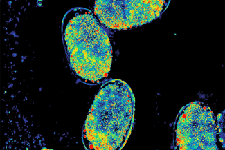
Computational microscopy enables imaging of optically scattering samples with rich label-free contrast. This picture differentiates C. elegans embryos by their developmental stages.
CZI's Frontiers of Imaging effort supports technology development to allow researchers to peer deep into tissues in order to better understand and cure disease.
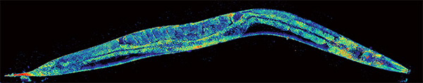
Computational microscopy can enable quantitative label-free imaging of multicellular organisms, which are often difficult to image due to strong optical scattering. This image of whole adult C. elegans worm can clearly and quantitatively visualize its internal anatomical components at sub-micron resolutions.
“Our goal is to support the advancement of imaging technologies and provide access to and training on these state-of-the-art tools so that researchers can drive towards discoveries,” said CZI Imaging Program Officer Stephani Otte. “By collaborating closely with the imaging community and providing both funding and expertise in technology development, we hope to help make the next breakthroughs in imaging possible.”
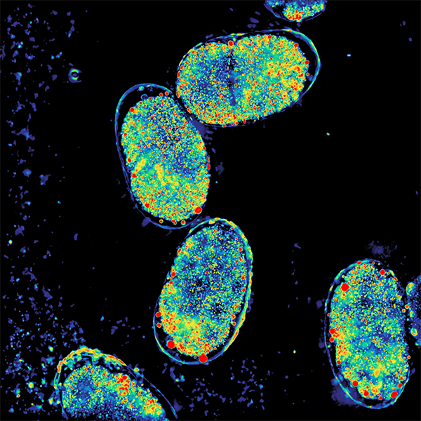
Computational microscopy also enables 3D volumetric reconstruction, which can help visualize internal 3D spatial distributions. 3D visualization of the collection of C. elegans embryos.
Shwetadwip Chowdhury will join Texas ECE in Spring 2021 as an assistant professor. His research interests are in developing next generation optical imaging technologies for applications in science and medicine. A key emphasis in his work is the joint design of novel optical imaging systems and advanced computational frameworks. This co-design of hardware and software enables imaging capabilities not possible in traditional optical imaging systems.
Previously, he was a NIH Ruth L. Kirschstein NRSA Postdoctoral Fellow at University of California Berkeley, in the Department of Electrical Engineering and Computer Sciences. Before that, he received his Ph.D. and B.S. degrees from the Department of Biomedical Engineering at Duke University.
Laura Waller is an associate professor in the Department of Electrical Engineering and Computer Sciences at the University of California, Berkeley, where she leads the Computational Imaging Lab.
